Liposomal Characterization of Malvern Instruments for Drug Delivery Systems
Author: Pauline Carnell, Malvern Instruments Senior Application Scientist; Mike Kazsuba, Malvern Instruments Technical Support Manager
Image: 4 in total (only available at the end of the file, high resolution image files are available separately)
Liposomal Characterization for Drug Delivery Systems
Malvin Instruments' senior application scientist Pauline Carnell and technical support manager Mike Kazsuba explored the application and effectiveness of nanoparticle tracking and analysis techniques and light scattering techniques in characterizing liposomes as drug carriers.
Liposomes are an important delivery vehicle and have been approved for use in a variety of therapeutic formulations. Liposomes are composed of phospholipids, have a single layer or a multi-layer structure, have a hydrophilic inner layer and a hydrophobic outer layer, and can be made into particles of different sizes. These particles are biodegradable and substantially non-toxic. Most importantly, it encapsulates both hydrophilic and hydrophobic materials. In addition, by modifying the surface of the liposome, it is also possible to target the specific physiological site, prolong the retention time of the liposome in the body, and can be used to design a diagnostic tool.
As with other similar studies, the key to applying liposomes is to ensure that their physical properties are consistent with their use. For example, how do liposomes react when they enter the body? Is the liposome stable enough to ensure targeting? Is the particle size suitable for clinical use, or will it disappear in the blood circulation?
Understanding the particle size, concentration, and zeta potential of liposome preparations can help predict its tendency to change in vivo, and the molecular relationship between charged liposomes and opposite electrical properties can also be measured by measuring the zeta potential of the polymer produced by both. monitor. These factors have a significant impact on the effectiveness of drug delivery, especially when a drug formulator believes that a liposome is suitable for delivery vehicles. Therefore, an analytical system that provides comprehensive data can be of great benefit to the formulation process. Nanoparticle tracking analysis technology and dynamic light scattering technology are two important analytical methods that provide important information for liposome research.
Nanoparticle tracking analysis
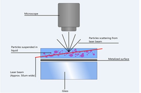
Nanoparticle Tracking Analysis (NTA) uses laser light scattering to verify nanoparticle size in solution (Figure 1). Using this analytical method, the researchers were able to observe individual particles and track their Brownian motion trajectories, so that the particle size distribution of each particle was quickly produced based on individual particles in a short period of time.
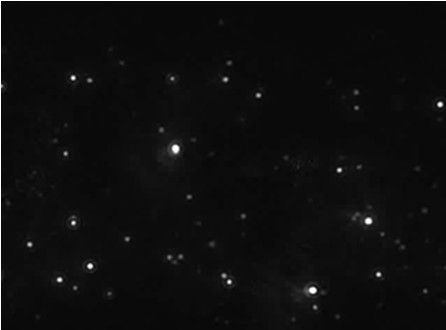
The scientific digital camera captures the scattered light from the particles in the solution, and the instrument software tracks the trajectory of each particle on a frame-by-frame basis (Figure 2).
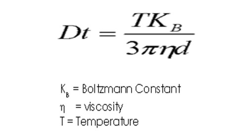
The velocity of the particles is related to the equivalent hydrodynamic radius of the sphere calculated by the Stokes-Einstein equation (Fig. 3). NTA technology can calculate granularity on a grain-by-grain basis, and users can accurately characterize real-time dynamics based on the analysis of image segments.
NTA technology allows researchers to view individual nanoparticles at the same time, so in addition to the basic particle size analysis, the relative light scattering intensity of each liposome can be determined. By plotting the data results with separately measured particle size data into a graph, the particles composed of different refractive indices (RI) or materials can be more carefully distinguished. With this unique feature, researchers can explore whether the content encapsulated by nanoscale drug delivery vehicles (such as liposomes) is different: the refractive index (light scattering ability) of hollow liposomes may be lower than that of higher refractive index Rate the substance of the liposome. This difference allows one to distinguish between liposomes of similar size. In addition, NTA's single particle detection system makes particle concentration measurement possible.
Particle size and zeta potential
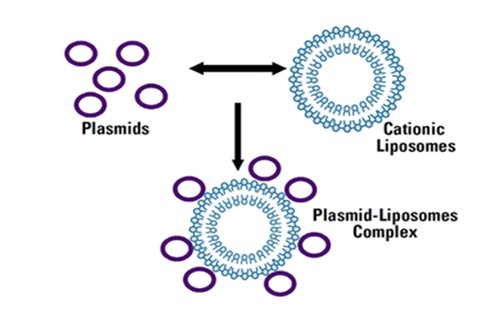
The location of action of liposomes and cells in vivo is largely determined by the particle size of the liposomes. Mastering the zeta potential of liposomal formulations helps predict the trend of liposomes in vivo. The zeta potential of a particle refers to the total charge that the particle acquires in a particular medium. In the case of gene therapy, zeta potential measurements can be used to optimize the ratio of specific liposomes to various DNA plasmids to minimize formulation aggregation (Figure 4).
Dynamic Light Scattering (DLS) is a relatively mature and widely used liposome characterization technique. In addition, since zeta potential is also an important parameter, an analysis system capable of simultaneously measuring particle size and zeta potential is becoming more and more popular, and Malvern Instruments' Zetasizer Nano system is one of them. In general, researchers used dynamic light scattering to measure particle size and laser Doppler microelectrophoresis to measure zeta potential.
Light scattering by the Brownian motion of the particles is also at the heart of the DLS technology. The DLS technique measures the fluctuations in the intensity of the scattered light over time and determines the diffusion coefficient of the particles. Based on this, the Stokes-Einstein equation is used to transform the data into a particle size distribution.
When the zeta potential is measured using laser Doppler microelectrophoresis, an electric field is applied to the molecular solution or particle dispersion, and the particles move at a rate which is related to the zeta potential. The electrophoretic mobility can be calculated by measuring the rate, and the zeta potential and the zeta potential distribution of the particles are calculated accordingly.
in conclusion
The physical characterization of liposomes is important for understanding the suitability of liposomes in a variety of applications, and rapid, reproducible characterization is an important consideration in R&D and quality control processes. The techniques described herein provide additional information such as particle size, concentration, and zeta potential of liposomal formulations. (End)
Caption: (for reference only - HD pictures are available separately)

Figure 1: Nanoparticle tracking analysis technology effect display

Figure 2: The light spot in the figure is the particle in Brownian motion

Figure 3: Stokes-Einstein equation

Figure 4: Complexation of cationic liposomes (positively charged) with DNA (plasmid)
Care Tools,Skin Care Tools,Acne Removal Tools,Acne Removal And Care Tools
Shenzhen Sipimo Technology Co., Ltd , https://www.sipimotech.com
